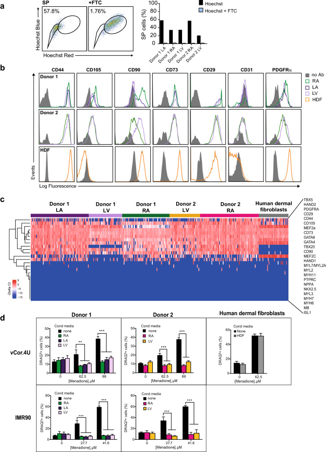
Figure 5.
Human cardiac stromal cells protect human cardiomyocytes. (a) Human cardiac stromal cells are enriched for the SP phenotype. Left, representative dot plots of SP staining for the left atrium of Donor 1. FTC, ABCG2 inhibitor Fumitremorgin C. Right, bar graphs showing consistent enrichment for SP cells in five human cardiac stromal cell populations from two donors. (b) Flow cytometry comparing surface marker expression in the human cardiac stromal cell populations and human dermal fibroblasts (HDFs). Note the absence of CD105 in HDFs. (c) Single-cell qRT–PCR comparing the co-expression of selected genes in human cardiac stromal cells and HDFs. The heatmap shows expression as − ΔCt values (blue, low; red, high). Genes are ordered based on hierarchical clustering. n = 39–72 cells for each sample illustrated. See also Fig. S3 in the supplementary data. (d) Human cardiac stromal cells from the donors and chambers shown all suppress cell death induced by menadione in vCor.4U (top) and IMR-90 (bottom) human cardiomyocytes; HDFs (right) had no effect. Bar graph, mean ± SEM; vCor,4U: n = 6; IMR-90: n = 9; **p < 0.001; ***p < 0.0001. RA right atrium, LA left atrium, LV left ventricle.

