Figure 6.
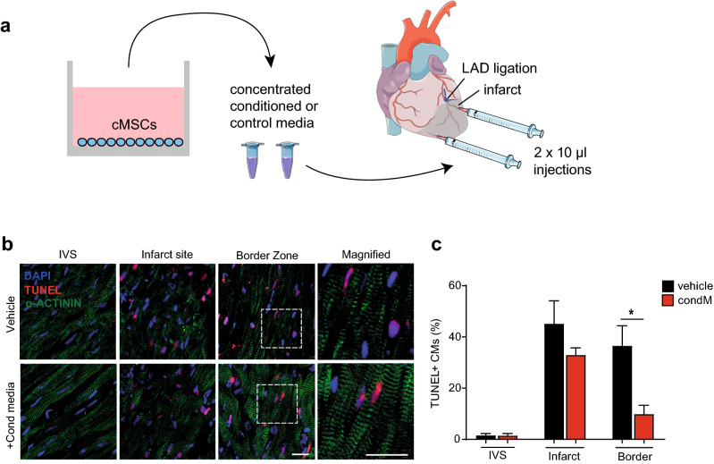
Conditioned media from mouse cMSC suppress cardiomyocyte apoptosis after mouse myocardial infarction. (a) Schematic representation of cMSC-conditioned medium production, concentration and intramyocardial injection after LAD ligation. Concentrated conditioned or control media were injected into the infarct border zone, one site ~ 1 mm beneath the suture and a second site more apical. Images modified from Servier Medical Art website, a free medical image database with a licence under Creative Commons Attribution 3.0 Unported License (https://creativecommons.org/licenses/by/3.0/). (b) Representative immunohistochemistry images of TUNEL staining, 24 h after myocardial infarction. An α-actinin antibody was used to demarcate cardiomyocyte identity. Scale bars 20 μm. See also Supplementary Fig. S11. (c) Bar graph of TUNEL staining in cardiomyocytes in the remote myocardium (intraventricular septum, IVS), infarct site, and border zone. Data are shown as the mean ± SEM. n = 3; *p < 0.05.

