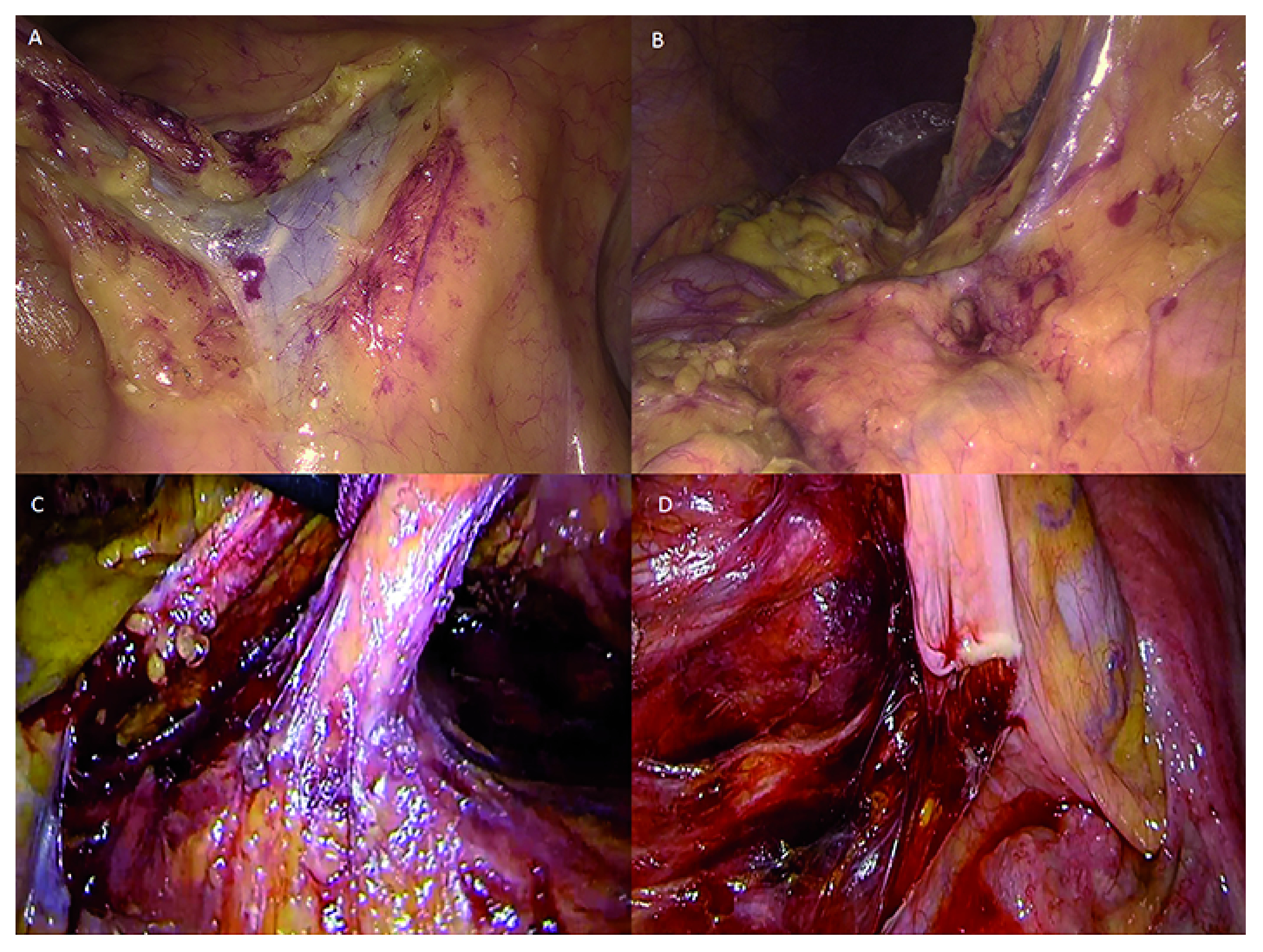
Figure 1.

A) Ileocolic vein and inferior mesenteric vein (HD technology). B) Middle colic vein (4K ultra HD technology). C) Gerota’s fascia and inferior mesenteric artery (HD technology). D) Hypogastric plexus at the origin of the inferior mesenteric artery (4K ultra HD technology). Images were obtained retrospectively from the video electronic library of our institution, which contains records of all minimally invasive procedures of patients who gave consent for publication of the images for educational or research purposes.
