Figure 2.
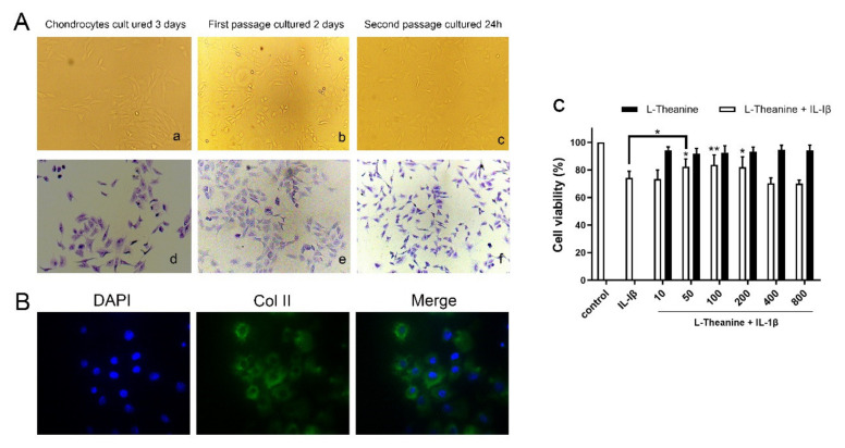
Identification of primary chondrocytes and the effects of L-theanine on cell viability with or without IL-1β. (A) Representative images of rat primary chondrocyte morphology, which was observed on the third day after incubated, the second day after the first passage of cells and 24 h after the second passage of cells, respectively(a-c). Representative images of toluidine blue staining of monolayers of rat chondrocytes in the same time point (d–f) and the nucleus of cells was dyed blue violet. Original magnification × 100. (B) Immunofluorescence assay of rat second generation chondrocytes for type II collagen with primary antibody to COL2A1 and fluorescent secondary antibody. DAPI for nucleus staining. Original magnification × 200. (C) CCK-8 assay for cell ability of L-theanine with or without IL-1β. L-theanine treated with different concentrations (0, 10, 50, 100, 200 400, 800 μM) showed no significant difference compared to controls. Pre-treatment with IL-1β (10 ng/mL) for 24 h sharply reduced cell viability, however the significant increase was observed after treatment of L-theanine in 50, 100, and 200 μM for 24 h. Values are the mean ± standard deviation (SD); * p < 0.05, ** p < 0.01 vs. IL-1β group.

