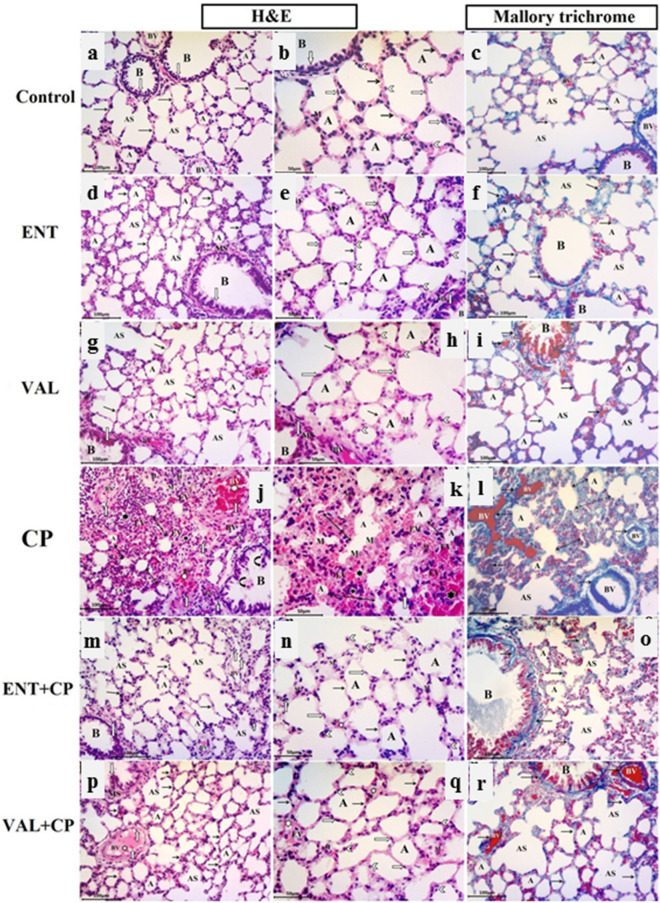
Figure 6.
Photomicrographs of lung sections from all experimental groups. ENT (sacubitril/valsartan), CP (cyclophosphamide), VAL (valsartan). Control group (a–c): (a) normal alveoli (A) separated by thin interalveolar septa (black arrow), alveolar sacs (AS), A patent bronchiole (B) , pulmonary blood vessels (BV). (b) Squamous type I pneumocyte with flattened nuclei (transverse white arrow) and cuboidal type II pneumocytes with rounded nuclei (white arrow head) bulging into the alveolar lumen. A few macrophages (M) in the interalveolar septa. Bronchiole (B) with simple columnar epithelium (vertical white arrow) and smooth muscle layer (SM). (c) Fine collagen fiber in alveolar sacs (AS) and in the thin interalveolar septa (black arrow) in between alveoli (A), around bronchiole (B) and blood vessels (BV). ENT and VAL groups (d–i): The normal alveolar architecture and collagen distribution appear as the control. CP-induced group (200 mg/kg; i.p.) single dose on day 5 (j–l): (j, k) narrowed alveolar spaces (A), thick interalveolar septa (double head arrow) , diffuse lymphocyte infiltration (black star), extravasated RBCs (EV), thick congested pulmonary blood vessels (white star) with lymphocytic infiltration (white arrow). Desquamated cells (curved arrows) in the bronchiolar lumen (B). (k) Large alveolar macrophages (M) with vaculated acidophilic cytoplasm in the pulmonary interstitium and within alveolar lumen. (l): Excessive collagen fiber deposition in thick interalveolar septa (detached double head arrow) and around congested pulmonary blood vessel (detached arrows). ENT + CP (30 mg/kg; p.o. for 6 days and 200 mg/kg; i.p. single dose on day 5, respectively) and VAL + CP groups (15 mg/kg; p.o. for 6 days and 200 mg/kg; i.p. single dose on day 5, respectively) (m–r): show a potentially alleviated lung architecture. Thickening of interalveolar septa in some regions (black arrow) with congested intervening blood vessels (white star) and a few extravasated RBCs (EV). (o, r) Normal distribution of collagen fibers in the relatively thin interalveolar septa (arrows), in between alveoli (A) , alveolar sacs (AS) , around bronchioles (B) and pulmonary blood vessels (BV) (H & E stain, X200, X400) and (Mallory trichrome stain, X200).

