
Fig. 2.
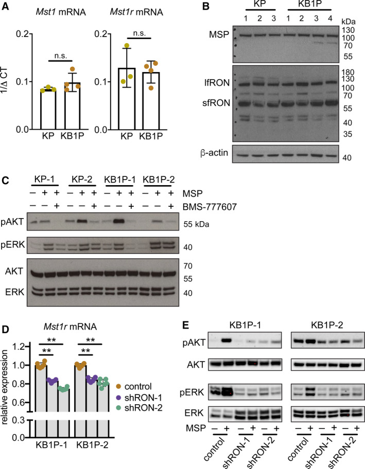
MSP stimulates AKT and MAPK signaling pathways through the RON receptor. (A) Mst1r mRNA expression assessed by qRT–PCR in cell lines derived from three KP tumors and four KB1P tumors, respectively. (B) Western blot analysis of RON protein expression in the same cell lines used in A. β‐Actin was used as sample integrity control (same sample, different blot). (C) Western blot analysis of indicated proteins in the same KP and KB1P cell lines used in A, B. Cells were pretreated with 1 μm BMS‐777607 for 1 h prior to 100 ng·mL−1 recombinant MSP for 1 h where denoted. Images are representative of three replicate experiments. Total AKT and ERK were probed on the same blot as sample integrity controls. (D) Two KB1P cell lines were transduced with lentiviral shRNA vectors against Mst1r mRNA (shRON‐1 or shRON‐2) or control pLKO.1 vector. Confirmation of Mst1r mRNA knockdown quantified by qRT–PCR is expressed as relative to two housekeeping genes, Hprt and β‐actin (each dot represents one technical replicate in a given experiment, the experiment was repeated three times for each cell line). (E) Two independent KB1P cell lines transduced with control or shRNA vectors against Mst1r mRNA were treated with 100 ng·mL−1 recombinant MSP for 1 h. Activation of AKT and ERK1/2 was assessed by western blot. Images are representative of three replicate transduction experiments. Data are represented as mean ± SD. **P < 0.01 as determined by one‐way ANOVA followed by Dunn’s post hoc test. n.s., not significant.