
Fig. 2.
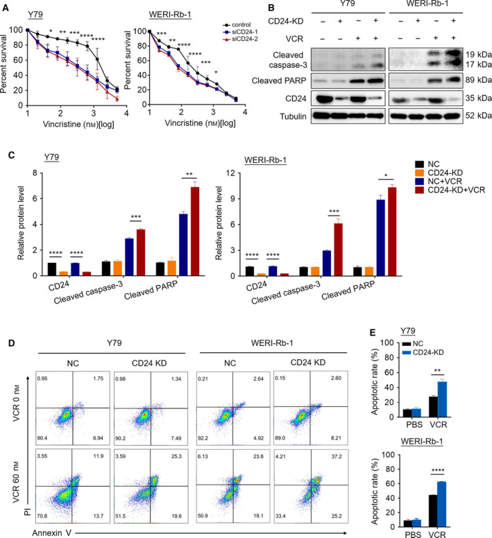
CD24 impedes the sensitivity of RB cells to VCR. (A) The CCK‐8 assay was performed in CD24‐silenced RB cell lines and their nontarget control cells after exposure to a serial dose–response of VCR for 48 h (n = 3, Student's t‐test). (B) Western blotting was performed in CD24 knockdown (CD24 KD) and control RB cells treated with PBS or VCR (60 nm) for 48 h. (C) Quantification analysis of the western blot image shown in (B) using imagej software (National Institutes of Health, Bethesda, MD, USA). (D, E) Fluorescence‐activated cell sorting (FACS) showing the apoptotic rate of cells stained with Annexin V‐APC/PI following 48 h of treatment with PBS or VCR. Representative plots (D) and quantification (E) are shown (n = 3, Student's t‐test). Quantitative data are presented as mean ± SD (*P < 0.05, **P < 0.01, ***P < 0.001, ****P < 0.0001).