
Fig. 4.
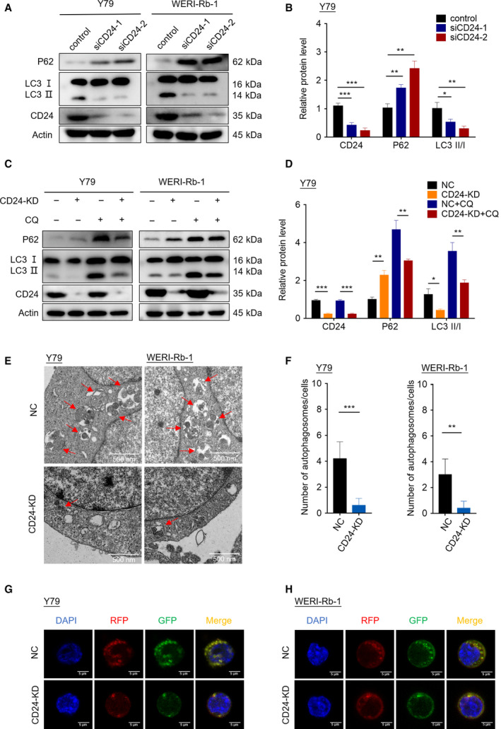
CD24 regulates autophagy activation in RB cells under VCR challenge. (A) The expression of LC3B and p62 protein was analyzed by western blotting. (B) The western blot images of Y79 cells in (A) were quantitatively analyzed using imagej software (National Institutes of Health, Bethesda, MD, USA). (C) Western blotting was performed in RB cells with or without CQ (20 μm) treatment for 1 h. (D) The western blot images of Y79 cells in (C) were quantitatively analyzed using imagej software. (E) Autophagosomes were observed by transmission electron microscopy in CD24 KD and control RB cells treated with CQ for 1 h. Scale bars, 0.5 μm. (F) Statistical analysis was performed to calculate the number of autophagosomes in RB cells shown by transmission electron microscopy (n = 5, Student's t‐test). (G, H) Y79 cells (G) and WERI‐Rb‐1 cells (H) that stably expressed RFP‐GFP‐LC3 fusion protein were treated with CQ for 1 h. The fluorescence signals were visualized by confocal immunofluorescence microscopy. Scale bars, 5 μm. Quantitative data are presented as mean ± SD (**P < 0.01, ***P < 0.001).