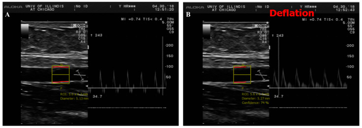
Figure 1.
Duplex B-mode/pulsed wave Doppler (PWD) ultrasound of brachial artery flow-mediated dilation (FMD). This figure shows a long axis scan of brachial artery with simultaneous blood velocity profile by pulsed wave Doppler before (A) and after cuff deflation (B). For offline image analyses, a representative section of brachial artery is selected for an automated measurement of diameter. Baseline diameter (BSL) was averaged from a serial of recorded frames before cuff deflation and the maximum diameter was measured after cuff deflation during reactive hyperemia (RH).

