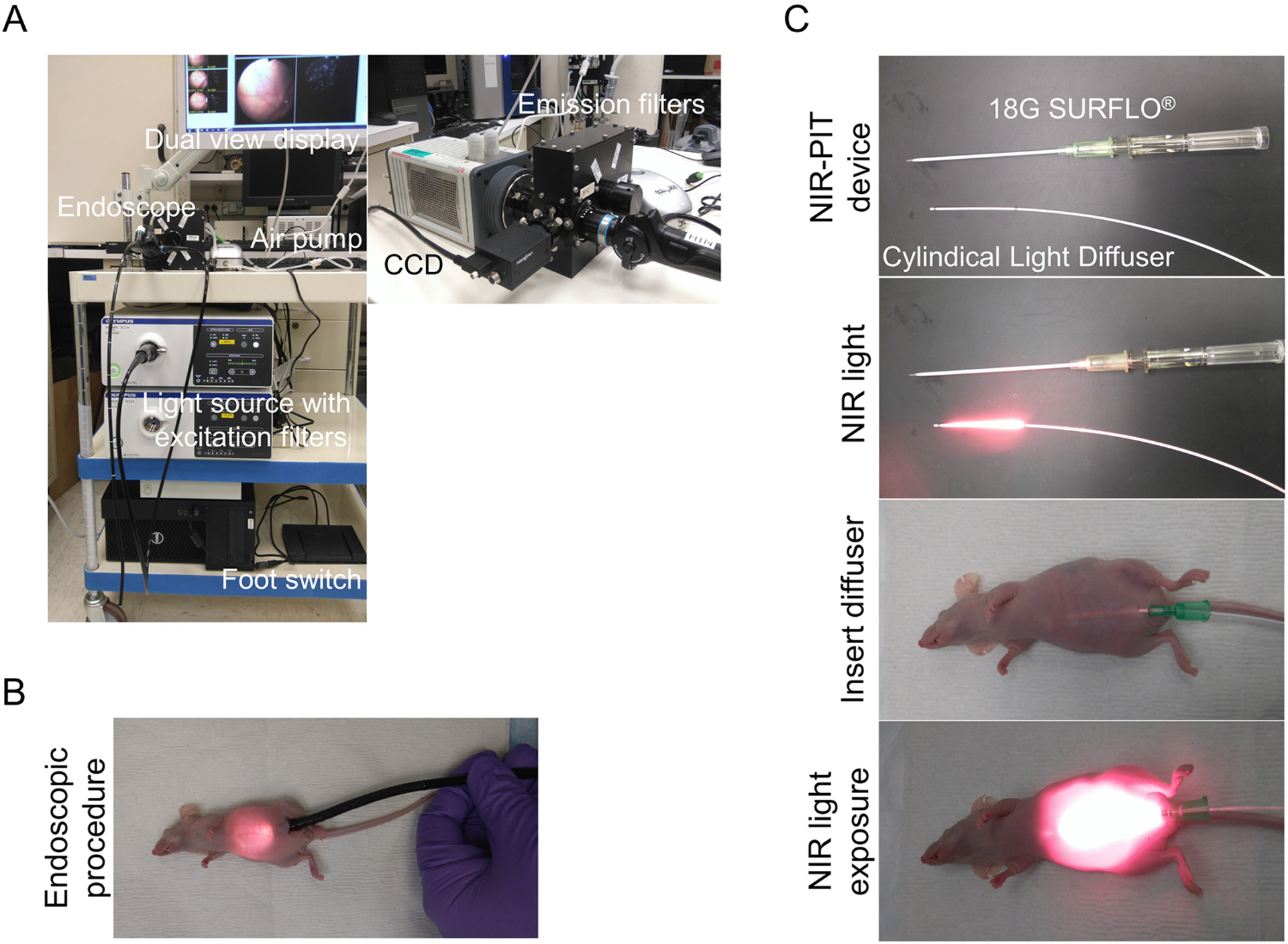
Figure 1. Multi-color fluorescence mini-endoscopic system and NIR-PIT procedure.

(A) Multi-color fluorescence imaging system is based on a clinically available fiber optic endoscope and light source. Excitation light is provided by multi-band excitation filters. Endoscopic images were obtained via a beam splitter, where the white light images were detected using the Color-CCD camera and the fluorescence images were filtered by multi-color emission filters and detected with an (EM)-CCD camera. Both images are displayed side by side on the monitor. (B) The endoscope was inserted into abdominal cavity through a small lower abdominal incision, and the abdominal cavity was inflated with air. (C) Peritoneal cavity was exposed to NIR light using a Cylindical Light Diffuser with a NIR laser through the 18G × 2 1/2” SURFLO®.
