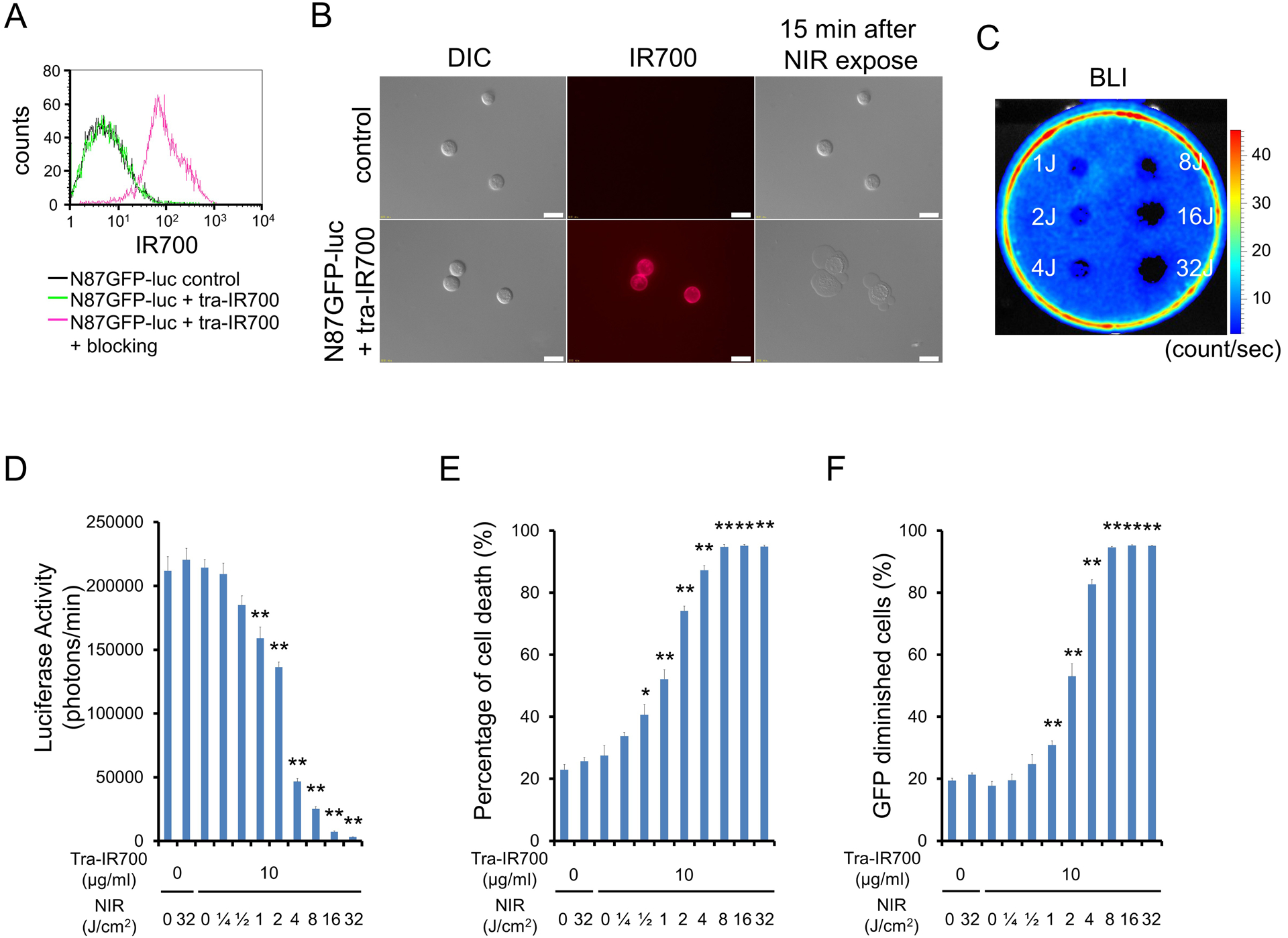
Figure 2. Confirmation of HER2 expression as a target for NIR-PIT in N87GFP-luc cells, and evaluation of in vitro NIR-PIT.

(A) Expression of HER2 in N87GFP-luc cells was examined with FACS. After 6 h of tra-IR700 incubation, N87GFP-luc cells showed high fluorescence signal. (B) Differential interference contrast (DIC) and fluorescence microscopy images of N87GFP-luc cells. High IR700 fluorescence signal was shown in N87GFP-luc cells after incubation with tra-IR700 for 6 h. On the other hand, there was no fluorescence signal in N87GFP-luc cells without incubation with tra-IR700. Immunogenic/necrotic cell death was observed upon excitation with NIR light (after 15min) only in N87GFP-luc cells incubated with tra-IR700. Scale bars = 20 μm. (C) Bioluminescence imaging (BLI) of a 10 cm dish demonstrated that luciferase activity in N87GFP-luc cells decreased in a NIR-light dose-dependent manner. (D) Luciferase activity in N87GFP-luc cells was measured, which also decreased in a NIR-light dose-dependent manner (n = 5, **p < 0.01 vs. untreated control, by Student’s t test). (E) Membrane damage of N87GFP-luc cells induced by NIR-PIT was measured with propidium iodide (PI) staining, which increased in a light dose dependent manner (n = 5, *p < 0.05, **p < 0.01, vs. untreated control, by Student’s t test). (F) Diminishing GFP fluorescence intensity in N87GFP-luc cells after NIR-PIT was measured by FACS. The GFP diminished within cells in a light dose-dependent manner (n = 5, **p < 0.01, vs. untreated control, by Student’s t test).
