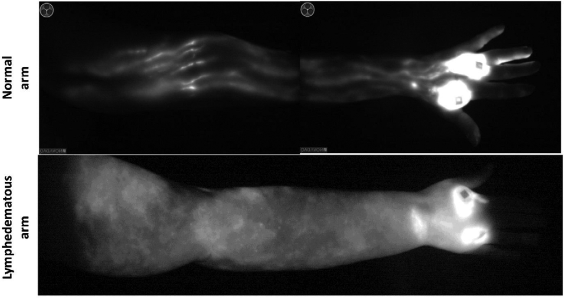
Figure 3: Indocyanine green (ICG) lymphangiography of a normal (upper) and lymphedematous (lower) upper extremity.

Note linear lymphatic collectors draining from the hand (bright spots are the injection sites) up towards the axilla in the normal limb. In contrast, note complete absence of lymphatic channels and accumulation of ICG dye in the skin of the lymphedematous limb.
