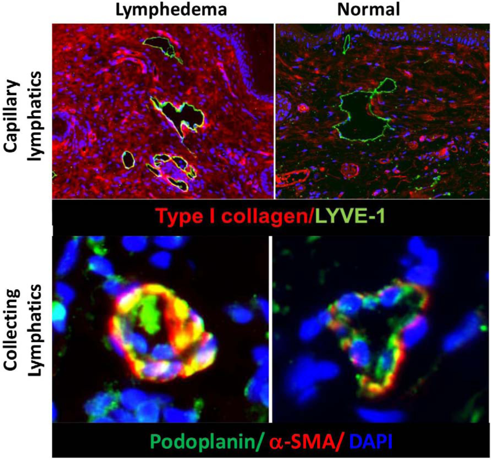
Figure 4: Lymphedema results in collagen deposition and fibrosis of capillary (upper panel) and collecting (lower panel) lymphatics in a mouse model of tail lymphedema.

Note accumulation of type I collagen fibers encasing capillary lymphatics in the skin (upper left). Also note proliferation of a-sma positive smooth muscle cells and obliteration of the lumen of collecting lymphatics in lymphedematous tissues (lower left).
