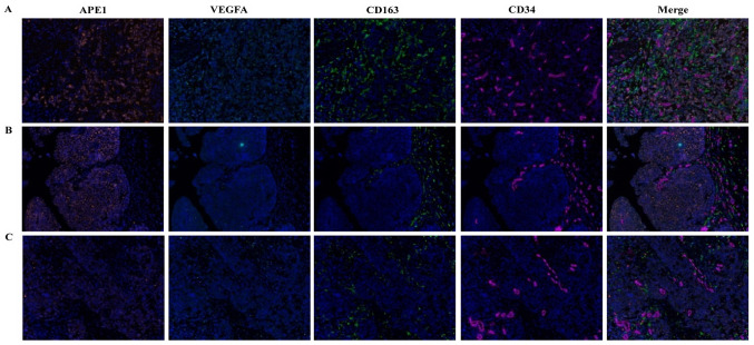
Figure 2.
Multiple immunofluorescence staining for APE1, VEGFA, CD163 and CD34 in bladder cancers. (A) Representative sample with high expression of APE1 and VEGFA in which the intratumor CD163-labeled M2 macrophages are easily distinguishable. (B) Example of a sample with high expression of APE1 and VEGFA in which the CD163-labeled M2 macrophages are easily distinguishable in the tumor stroma. (C) Representative sample with low expression of APE1 and VEGFA, in which the CD163-labeled M2 macrophages are less distinguishable. APE1-positive staining in tumor epithelial cells displayed as orange and VEGFA-positive staining in tumor epithelial cells was cyan, while CD163-positive infiltrating M2 macrophages were green and CD34-positive vascular endothelial cells displayed as magenta (magnification, ×200). VEGFA, vascular endothelial growth factor A; APE1, apurinic/apyrimidinic endodeoxyribonuclease 1.

