Figure 1.
SMAX1-GFP Is Functional, but Not Detectable Unless an RGKT Motif Is Removed.
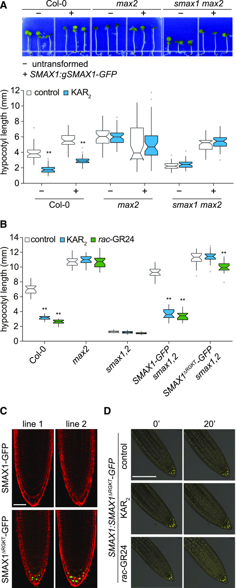
(A) Hypocotyl lengths of Col-0, max2-1, and smax1-2 max2-1 seedlings that are nontransgenic (–) or homozygous transgenic (+) for SMAX1:gSMAX1-GFP. Surface-sterilized seeds were plated on solid 0.5× MS medium, stratified 3 d at 4°C, given a germination initiation treatment (3 h of white light, 21 h of dark at 21°C), and grown under continuous red light (30 µE) for 4 d at 21°C before measurement. Boxplot notches approximate the 95% CI for the median. n = 45. *P < 0.01, **P < 0.001, two-way analysis of variance with Bonferroni’s test, comparisons to control treatment.
(B) Hypocotyl lengths of Col-0, max2-1, smax1-2 smxl2-1, and smax1-2 smxl2-1 lines homozygous for SMAX1:SMAX1-GFP or SMAX1:SMAX1∆RGKT-GFP transgenes. Assay conditions as described in (A), but medium was supplemented with 1 μM KAR2, 1 μM rac-GR24, or 0.1% (v/v) acetone as a solvent control. Boxplot notches approximate the 95% CI for the median. n = 20. *P < 0.01, **P < 0.001, two-way analysis of variance with Bonferroni’s test, comparisons to control treatment.
(C) Confocal images of 4-d light-grown seedlings from two independent transgenic lines (line 1 and line 2) for SMAX1:SMAX1-GFP or SMAX1:SMAX1∆RGKT-GFP in smax1 smxl2 background. Propidium iodide staining (red) was performed to visualize cell boundaries. Bar = 25 μm.
(D) Confocal microscopy of SMAX1:SMAX1∆RGKT-GFP smax1 smxl2 seedlings treated with 10 μM KAR2, 10 μM rac-GR24, and 0.1% (v/v) acetone control. Seedlings were grown vertically on 0.5× MS medium for 4 d under white light (∼150 μE, 12 h of light and 12 h of dark) following stratification. Images are overlays of bright field (gray) and GFP channel (yellow) images. Bar = 50 μm.

