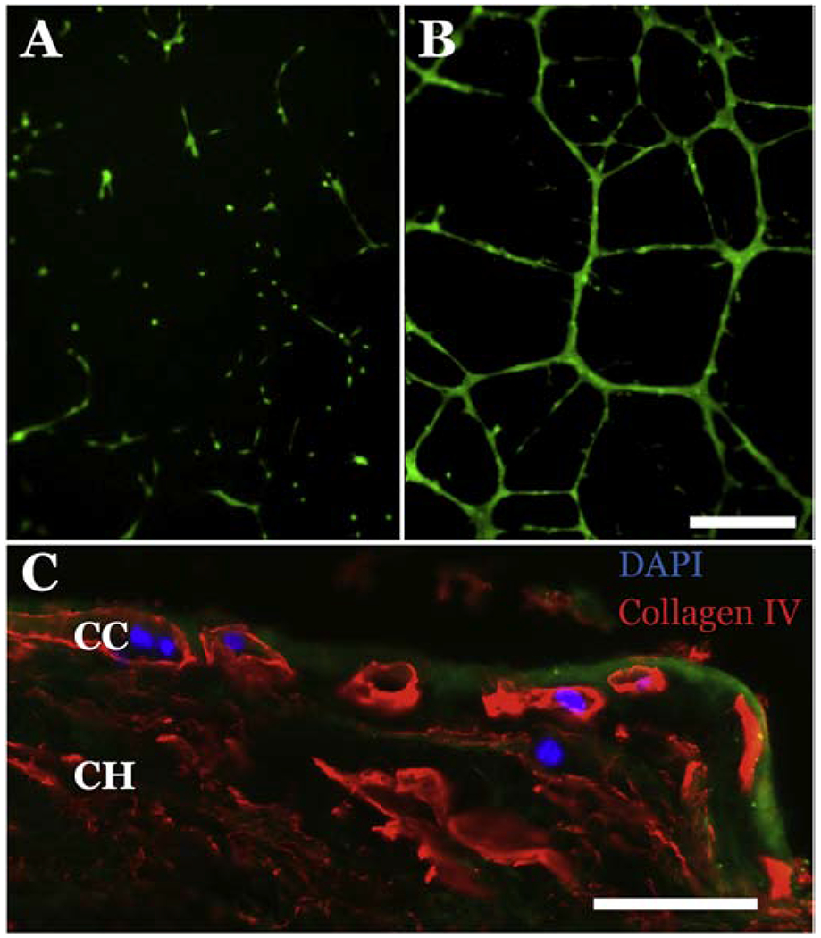
Figure 5. Assessment of iChEC-1 cell function.

A-B: Tube formation assay demonstrating that iChEC-1 cells form tubes in matrigel (A, 5×10⁴ cells; B, 1×10⁵ cells), note calcein-labeled (green) cells in vascular networks. Scalebar =250μm. C: iChEC-1 cells (nuclei, DAPI) home to empty capillary tubes (Collagen IV, red) in decellularized human choroid explants. Green = Phalloidin ; CC = choriocapillaris; CH= choroid, Scalebar = 50 μm.
