
Figure 1.
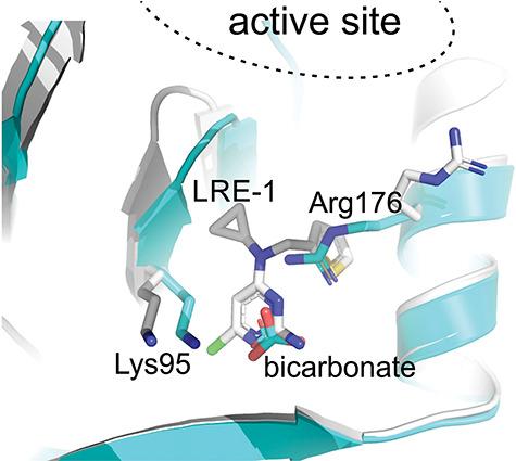
Crystal structures of ligands bound to sAC BBS. Crystal structure of bicarbonate in the sAC BBS (grey, PDB ID 4CLL), superimposed with the sAC − LRE1 complex (turquoise; PDB ID 5IV3). LRE1, bicarbonate, and the key bicarbonate-recognizing residues are shown in stick representation. The location of the active site where ATP binds is indicated [23, 46].