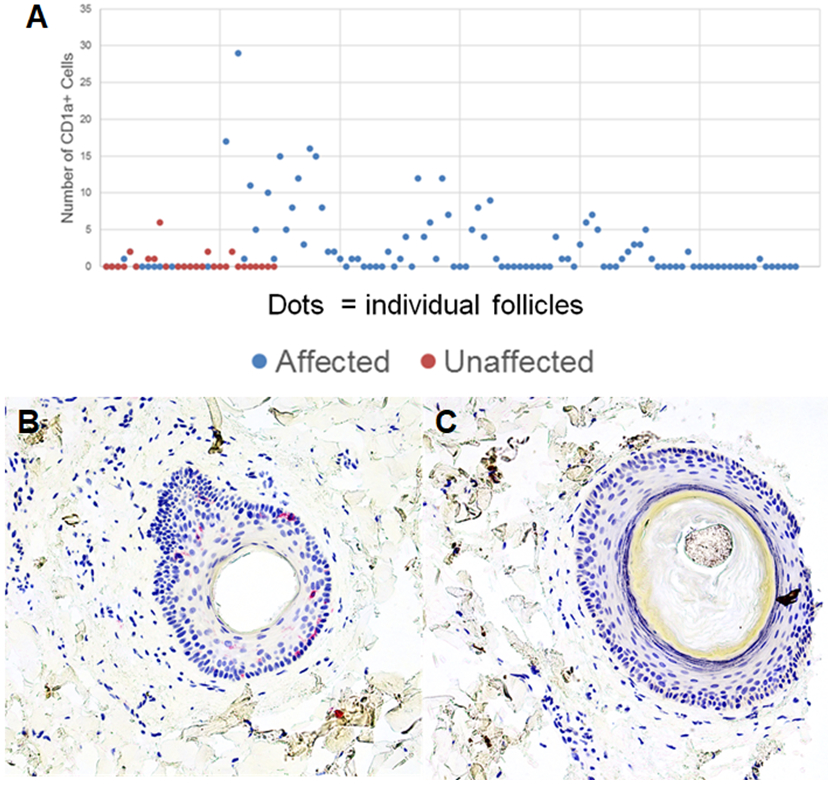
Figure 2: CD1a+ Cells Associated with Affected Follicles:

Affected and unaffected follicles were evaluated for the number of CD1a+ cells in the outer root sheath. Affected follicles have a higher number of CD1a+ cells than unaffected follicles (A). CD1a immunostaining of representative affected and unaffected follicle is seen in panels B and C, respectively. Photomicrographs 200x, B and C-hematoxylin.
