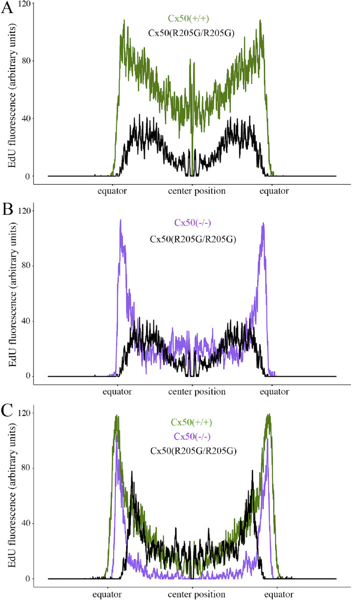
Figure 4.
Quantification of EdU labeling from the peripheral (equator/germinative zone) to the central epithelium of P2 and P3 lenses. (A) Line scans along the flattened EdU fluorescent images of P2 lenses, crossing through the diameter of the lens (a position of approximately 1500 µm is the center of the lens, and the position at which the fluorescence jumps from zero is the lens equator). The P2 Cx50(R205G/R205G) lens has lower epithelial cell proliferation by EdU labeling at all locations along the lens diameter compared with the P2 wild-type Cx50(+/+) (P < 0.01). (B) A comparison of line scans of EdU-labeled P2 Cx50(–/–) knockout and P2 Cx50(R205G/R205G) lenses. The P2 Cx50(R205G/R205G) lens has significantly lower proliferation in both central and peripheral (germinative zone) epithelium compared with the P2 Cx50(–/–) (P < 0.01). (C) Line scan comparison of EdU-labeled P3 wild-type Cx50(+/+), Cx50(–/–), and Cx50(R205G/R205G) lenses. The P3 Cx50(R205G/R205G) lens has a surge in proliferation but still shows decreased proliferation in peripheral epithelium when compared with that of both wild-type and Cx50(–/–) (P < 0.01). For all line scan charts, about 154 to 166 line scans per sample were averaged for each age and genotype (n = 3–5); SEM ribbon not shown due to large sample size and small SE. Student’s t-tests for the central epithelium performed at positions 1250 to 1750 µm.

