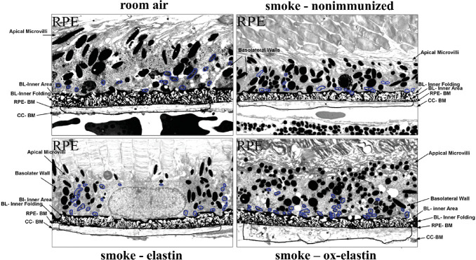
Figure 5.
Masks overlying the morphologic features to be analyzed. Electron micrographs described in Figure 4 were used for analysis. Adobe Photoshop was used to generate masks for BrM, basal infolding, and mitochondria for further analysis in ImageJ. Masks highlight the differences in RPE/BrM obtained from C57BL/6J mice exposed to 6 months of room air when compared with those exposed to 6 months of smoke in the absence (smoke – untreated) and presence of elastin immunization (smoke – elastin; smoke – ox-elastin).

