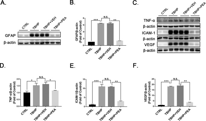
Figure 7.
Effects of PEA in TBHP-treated rMC-1 cells. Representative images of western blotting for (A) GFAP and (C) TNF-α, ICAM-1, and VEGF in Müller cells treated with VEH or PEA in the presence of TBHP. Protein levels of (B) GFAP, (D) TNF-α, (E) ICAM-1, and (F) VEGF were quantified by densitometry and normalized to β-actin levels. Data are presented as mean ± SEM; *P < 0.05, **P < 0.01, ***P < 0.001. TBHP, tertiary-butyl hydrogen peroxide.

