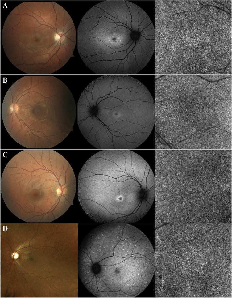
Figure 1.
Patients with early changes on SW-AF due to recessive STGD but healthy CC (group 1). Color fundus photography, SW-AF, and OCTA imaging of patients A–D. These patients present with a small area of macular hypoAF with a surrounding ring of hyperAF, both early changes associated with recessive STGD. Nevertheless, the CC appear healthy with no changes on OCTA imaging. Fundoscopy is unremarkable for patients A–C. For patient D, hyperAF flecks on SW-AF and yellow flecks on fundoscopy are observed, both characteristic of STGD.

