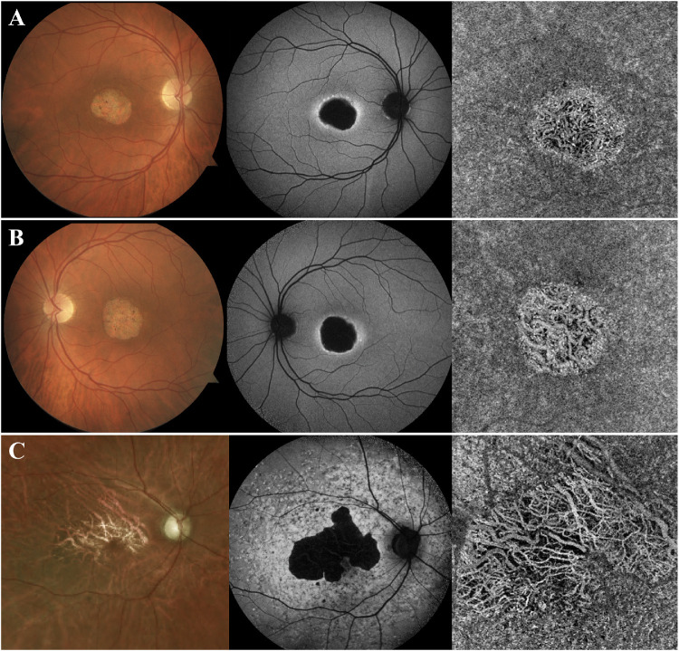
Figure 3.
Patients with STGD with extensive CC atrophy in areas of retinal atrophy (group 4). Patients A–C presented with extensive atrophy of the CC, with visualization of the underlying larger choroidal vessels. These areas of atrophy corresponded to areas of extensive RPE loss on SW-AF imaging, as suggested by the dense areas of hypoAF. Furthermore, the underlying white sclera was visible on fundoscopy on these areas.

