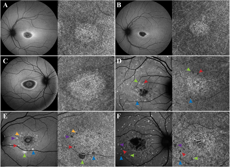
Figure 5.
Accumulation of lipofuscin in the overlying RPE creates a shadow on the underlying CC. In patients A–F, SW-AF reveals variable degrees of macular RPE atrophy, with the lesion appearing bright on the underlying CC as observed on OCTA imaging. This apparent brightness of the lesion is likely created as more signal is received on this area owing to the atrophy of the overlying RPE. In addition, an apparent dark halo is appreciated on the perimeter surrounding the bright lesion on the CC. This shadow is likely created due to signal blockage from the lipofuscin that has accumulated on the overlying RPE, as appreciated from the hyperAF ring surrounding the macular atrophy on SW-AF imaging. The blockage of signal by RPE lipofuscin is further supported by analyzing the hyperAF flecks in patients D–F, as these flecks create a dark spot on the CC images (indicated by the matching color arrows on SW-AF and OCTA imaging).

