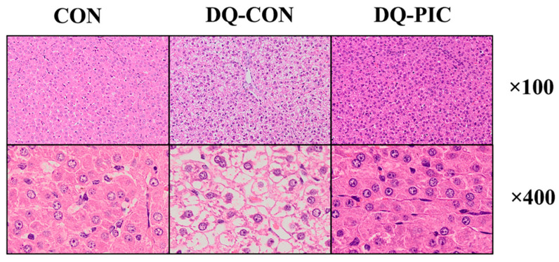
Figure 1.
Representative images of hepatic tissue assessed by hematoxylin and eosin staining. CON, piglets were orally administrated vehicle solution (0.5% sodium carboxymethyl cellulose) and challenged with sterile saline; DQ-CON, piglets were orally administrated vehicle solution and challenged with diquat (10 mg/kg body weight); DQ-PIC, piglets were orally administrated piceatannol (80 mg/kg/day) and challenged with diquat (10 mg/kg body weight).

