Figure 1.
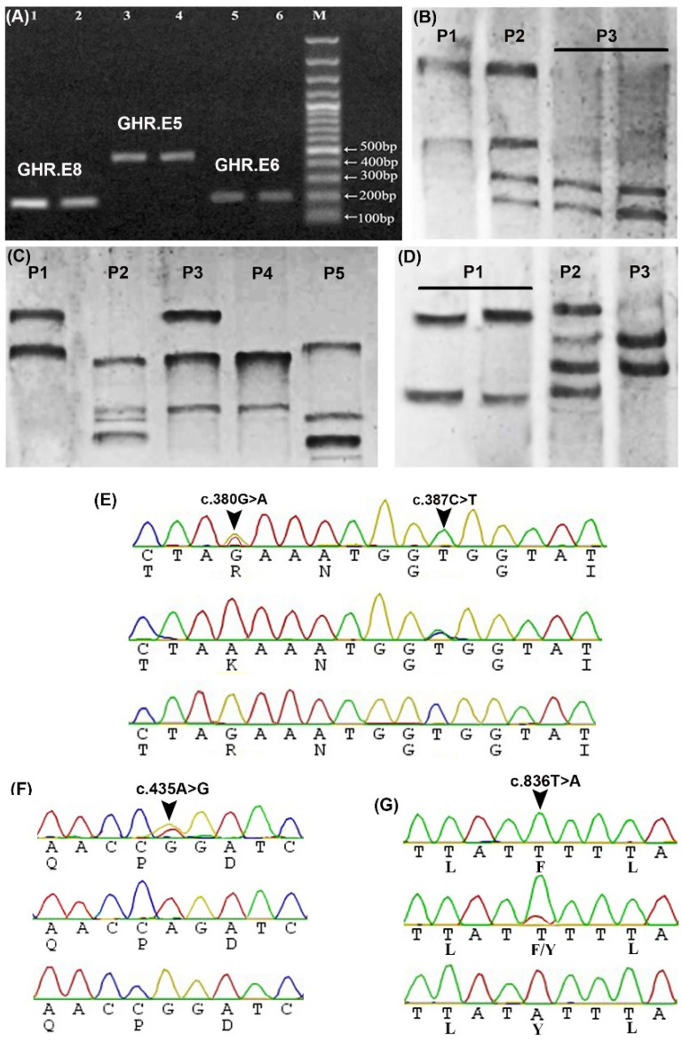
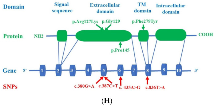
Detection of GHR SNPs using PCR- single strand conformation polymorphism (SSCP) and sequencing. (A) Agarose gel shows amplified fragments of GHR.E6 (205 bp, lanes 5 and 6), GHR.E5 (472 bp, lanes 3 and 4), and GHR.E8 (166 bp, lanes 1 and 2) from two different samples, M: 100 bp DNA ladder. (B) PCR-SSCP bands of GHR.E6 show three different patterns (P1-P3) in four different samples. (C) PCR-SSCP bands of GHR.E5 show five different patterns (P1-P5) in five different samples. (D) PCR-SSCP bands of GHR.E8 show three different patterns (P1-P3) in four different samples. (E) Sequences chromatogram spanning the site of c.380G>A (p.Arg(R)127Lys (K)) and c.387C>T (p.Gly(G)129) in E5. (F) Sequences chromatogram spanning the site of c.435A>G (p.Pro(P)145) in E6. (G) Sequences chromatogram spanning the site of c.836T>A (p.Phe(F)279Tyr(Y)) in E8. (H) Location of the predicted c.380G>A (p.Arg127Lys), c.387C>T (p.Gly129), c.435A>G (p.Pro145), and c.836T>A (p.Phe279Tyr) SNPs in the extracellular and transmembrane domains of the GHR protein. Blue cylinders indicate the exons (1–10).


