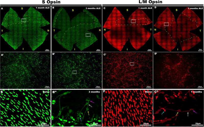
Figure 2.
Light exposure causes a disruption of the normal photoreceptor mosaic and morphological changes in the surviving photoreceptors. S- (left two columns, green) and L/M- (right two columns, red) cone immunodetection in whole mounts of the (A, B) left and (C, D) right retinas of four representative experimental animals, (A, C) 1 and (B, D) 3 months ALE. In the whole-mounted retinas, the dashed yellow lines surround, at 1 month ALE, the area of the superotemporal retina where the rings of (A) S- and (C) L/M-cone degeneration first appear and, at 3 months ALE (B, D), the areas devoid of S- and L/M cones. (A–D) Insets are shown at a higher power in A′–D′, respectively, to show the characteristic rings of photoreceptor degeneration. The insets in B′ and C′ are shown at higher power in B″ and C″ to illustrate the degenerating (B″) S and (C″) L/M cones at the periphery of the rings. Degenerating cones show opsin expression in the outer segments (blue arrow), somas (yellow arrow), inner segments (white arrow), axons and terminals (purple arrow), and their outer segments are located in the periphery of the rings, whereas their axons are directed towards the center of the rings.

