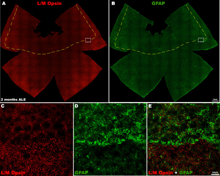
Figure 4.
L/M-cone loss and GFAP expression 2 months ALE. Photomontages of the same representative retinal whole mount of an animal processed two months ALE and immunoreacted for (A, red) L/M opsin and (B, green) GFAP showing that the superior “photosensitive area” of the rat retina is at this time point devoid of (A) L/M-cones, whereas there is an increased expression of (B) GFAP in this area and specially in its boundaries. (A, B) Insets are shown at higher power in (C) and (D), respectively, and (E) is a merged image of (C) and (D) to show the cone loss and increased GFAP immunoreactivity at the limits of the photosensitive area.

