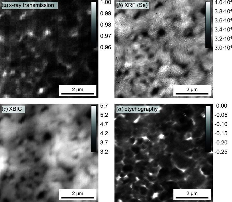
Figure 13.
Multi-modal X-ray imaging of a solar cell with a CuIn1−xGaxSe2 absorber layer. 2D maps of (a) the X-ray transmission signal (maximum intensity scaled to 1), (b) the XRF signal (sum of Kα and Kβ) of selenium (counts per second), (c) the XBIC signal (nanoamperes) and (d) the ptychographically reconstructed phase of the object transmission function (radians). See also the greyscale bars in each image.

