Figure 7.
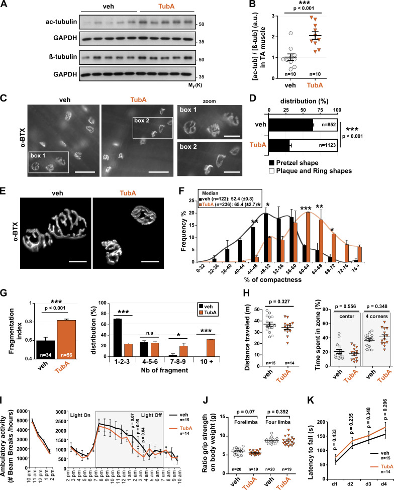
In vivo, HDAC6 inhibition via TubA treatment regulated NMJ structure and did not affect behavioral disorders. (A and B) 7-wk-old WT mice were treated with TubA or with vehicle-control (veh) for 31 consecutive days. To evaluate the level of α-tubulin acetylation in veh- and TubA-treated mice in TA muscles, Western blot analysis (A) and quantification (B) were performed. Quantification of acetylated tubulin protein level normalized to β-tubulin. GAPDH was used as a loading control (n = number of mice used per condition, 10). (C) Hemi-DIA muscle fibers were stained with α-BTX–A488 (in gray). (D) Total distribution between pretzel-like shape and plaque/ring shapes in hemi-DIA (n = total number of NMJs counted on five mice for each condition; veh = 852 and TubA = 1,123). (E) NMJs of isolated TA fibers labeled with α-BTX–A488 (in gray). (F) Graphical summary of NMJ compactness (n = total number of NMJs counted on five mice for each condition; veh = 122 and TubA = 236). (G) Fragmentation index and distribution of number of fragments have been quantified (n = total number of NMJs counted on three mice for each condition; veh = 34 and TubA = 56). (H) Open-field behavior. Distance traveled and time spent in the center or in corners of the open-field chamber are shown on the y axis. (I) Beam break test was realized for 12 h. Motor habituation and activity are shown on the y axis. (J) Grip strength was measured on a grid measuring maximal forelimb and hind limb grip strength normalized on body weight. (K) Rotarod test was performed on 4 d. Effect of TubA on the average time to fall off the rotarod. (H, I, J, and K) n = number of mice used per condition (veh = 15–20, and TubA = 14–19). Quantifications show means ± SEM. *, P < 0.05; **, P < 0.01; ***, P < 0.001; Mann-Whitney U test. Bars: 500 µm (C); 25 µm (E); inset magnifications (C; boxes 1, 2): 250 µm. n.s, not significant; β-tub, β-tubulin; Mr(K), relative molecular weight in kiloDalton; Nb, number; ac-tub and ac-tubulin, acetylated tubulin.

