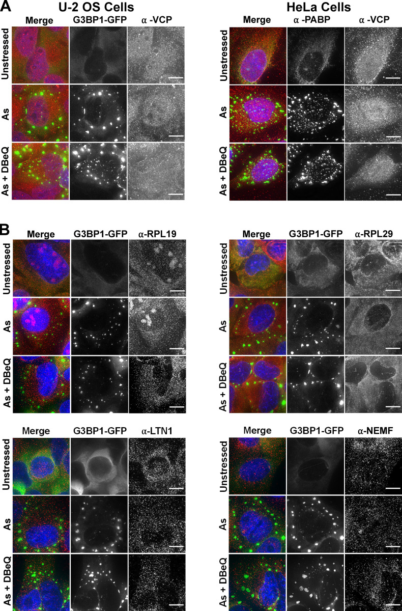
Figure S5.
VCP, LTN1, NEMF, and ribosomal proteins Rpl19 and Rpl29 are not enriched in SGs 45 min after arsenite stress. (A) U-2 OS cells stably transfected with the SG marker GFP-G3BP1 (green; left) or HeLa cells (right) were unstressed or stressed for 45 min with 0.5 mM sodium arsenite (As) in the presence or absence of DBeQ (10 µM). Cells were fixed, and IMF staining was performed to detect endogenous VCP (red); SGs in HeLa cells were visualized with anti–poly(A)-binding protein (PABP; green). Results represent n = three independent experiments. (B) U-2 OS cells expressing GFP-G3BP1 (green) were treated as described in A, and IMF staining was done to detect Rpl29, Rpl19, LTN1, or NEMF (red). Nuclei are shown in blue (DAPI). Cells were imaged at 100× with a DeltaVision elite microscope, and representative maximum intensity projections of 25 z-stacks are shown with 10-µm scale bars. Results represent n = two independent experiments.

