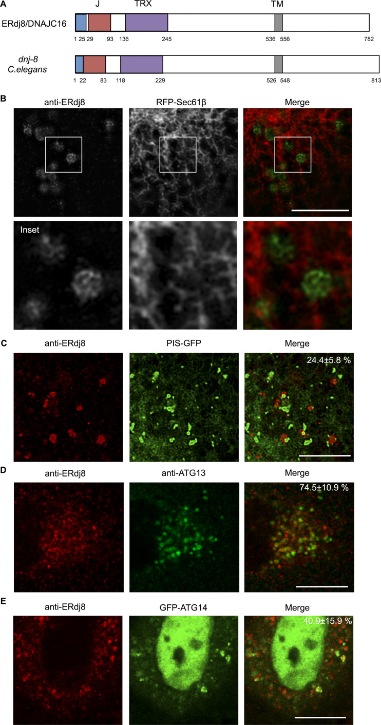
Figure 1.
ERdj8 is concentrated in ER subdomains. (A) Schematic diagram of ERdj8/DNAJC16 (Human) and dnj-8 (C. elegans). Blue, signal peptide; red, DnaJ domain (J); purple, thioredoxin-like domain (TRX); and gray, transmembrane region (TM). (B) COS-7 cells transfected with RFP-Sec61β were stained with anti-ERdj8 and imaged on SpinSR10. Insets, enlargements of framed regions. Scale bar, 10 µm. (C) COS-7 cells stably expressing PIS-GFP were starved for 2 h, stained with anti-ERdj8, and imaged on SpinSR10. Scale bar, 5 µm. The number is the percentage and SD of PIS-GFP–positive structures among ERdj8 structures per cell (n = 7). (D) HeLa cells were starved for 1 h, immunolabeled with ERdj8 and ATG13, and imaged on an SP-8. The number is the percentage and SD of ATG13-positive puncta among ERdj8 structures per cell (n = 10). Scale bar, 10 µm. (E) HeLa cells transfected with GFP-ATG14 were starved for 1 h, immunolabeled with ERdj8, and imaged on SP-8. The number is the percentage and SD of GFP-ATG14–positive structures among ERdj8 structures per cell (n = 10). Scale bar, 10 µm.

