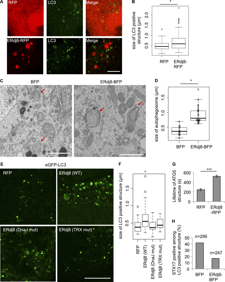
Figure 2.
Overexpression of ERdj8 increases autophagosome size. (A and B) HeLa cells were transfected with RFP or ERdj8-RFP, incubated under starved condition for 1 h, immunolabeled for LC3, and imaged on an SP-8. Scale bar, 10 µm. (B) Length of the most distal point in each of the LC3-positive structures. *, P < 0.05 by t test. *, P < 0.05 by ANOVA, Tukey–Kramer test. Mean of five cells ± SD. (C and D) HeLa cells stably expressing eGFP-LC3 were transfected with ERdj8-BFP or BFP and starved for 1 h. CLEM analysis was conducted. Scale bar, 1 µm. Red arrows show autophagosomes. (D) Length of the most distal point in each of the autophagosomes.Diameters of GFP-LC3–positive autophagosomes were measured in BFP (14 autophagosomes) and ERdj8-BFP (25 autophagosomes). *, P < 0.05 by t test. Mean of GFP-LC3–positive autophagosomes ± SD. (E and F) HeLa cells stably expressing eGFP-LC3 were transfected with ERdj8(WT)-RFP, ERdj8 (DnaJ muta)-RFP, ERdj8 (TRX mut)-RFP, or RFP only and imaged on an SP-8. Scale bar, 10 µm. (F) Length of the most distal point in each of the eGFP-positive puncta. *, P < 0.05 by ANOVA, Tukey Kramer test. Mean of five cells ± SD. (G) COS-7 cells stably expressing YFP-ATG5 were transfected with ERdj8-RFP or RFP and starved for 2 h. Live images of YFP-ATG5–positive structures were acquired on a DeltaVision system at intervals of 10 s. The average of 12 lifetimes of each YFP-ATG5 structure is shown. Mean of YFP-ATG5–positive puncta ± SEM. ***, P < 0.001. (H) HeLa cells stably expressing eGFP-LC3 were transfected with mCherry-STX17 and ERdj8-BFP or BFP only (as a control), starved for 2 h, and imaged on an SP-8. Percentage of mCherry-STX17–positive among GFP-LC3–positive structures. Total numbers of eGFP-LC3–positive structures counted are shown as n.

