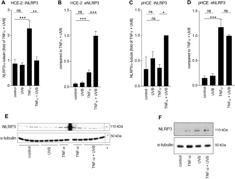
Figure 3.
Fate of NLRP3 in UV-B-stressed HCE cells. HCE-2 cells were primed for 24 hours with TNF-α (10 ng/mL) following irradiation with UV-B (0.2 J/cm2), and then incubated for 24 hours before sample collection and analysis. Intracellular levels of NLRP3 (iNLRP3; A) were measured by the Western blot technique from cell lysates and the levels of NLRP3 were normalized to those of α-tubulin. Data in panel A is combined from two (UV-B) to four (untreated control, TNF-α, TNF-α + UV-B the correct group is TNF-α + UVB. This means that extra red comma should be removed.) independent experiments with three to four parallel samples in each group. Extracellular levels of NLRP3 (eNLRP3; B) were measured by the ELISA method from HCE-2 cell culture supernatants and data were combined from three independent experiments with three parallel samples in each group. Experiments were repeated using primary HCE cells, and intracellular levels of NLRP3 were measured by Western blot from cells of four donors (C), and extracellular levels of NLRP3 by ELISA from four donors with two parallel samples (D). (E, F) Representative Western blots from NLRP3 and α-tubulin measurements of (A) HCE-2 cells lysates or (C) primary HCE cell lysates. All data are presented as mean ± SEM. *P < 0.05, **P < 0.01, ***P < 0.001, ns = not significant, Mann-Whitney U-test; + the NLRP3 positive control + equals to the NLRP3 positive control.

