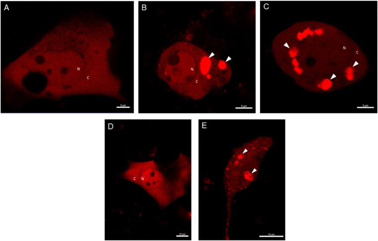
Figure 4.
The formation of ASC specks upon UV-B irradiation in HCE cells. HCE-2 cells were transfected with DsRed-ASC plasmid constructs for 24 hours and primed for the next 24 hours with TNF-α (10 ng/mL). HCE-2 cells were observed under the confocal microscope 5 hours after the UV-B exposure (0.2 J/cm2; A = control group and B-C = TNF-α + UV-B treated group). Alternatively, microscopic examinations were performed 24 hours after the UV-B irradiation (D = control group and E = TNF-α + UV-B treated group). Experiments were repeated three times and images were photographed using the 63-fold objective (A-C: scale bar, 5 µm; D-E: scale bar, 10 µm). White arrows indicate ASC specks. N = nucleus, C = cytoplasm.

