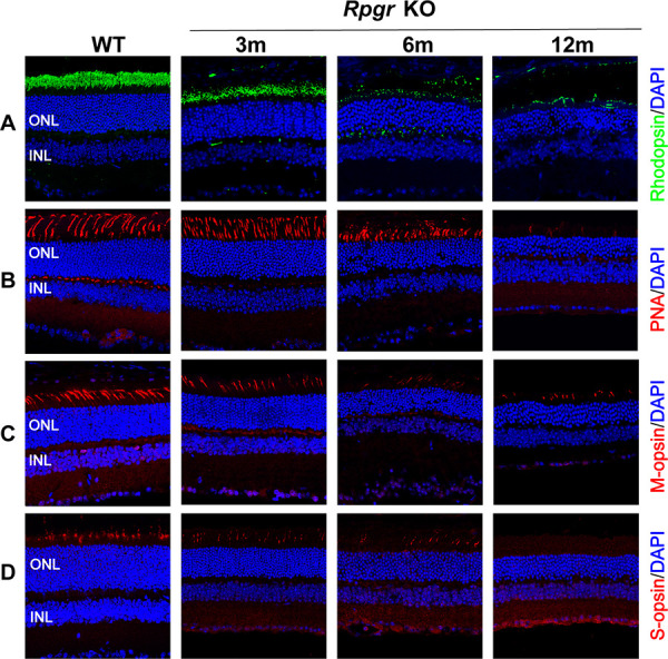
Figure 3.

Photoreceptor degeneration in Rpgr KO mice. Immunofluorescent staining of rhodopsin (A), PNA (B), M-cone opsin (C), and S-cone opsin (D) in Rpgr KO mice. All staining was substantially reduced in 12-month-old Rpgr KO mice. PNA, M-cone, and S-cone staining are shown in red. Rhodopsin staining is shown in green. Nuclei are stained blue by DAPI. Scale bar: 50 µm. INL, inner nuclear layer.
