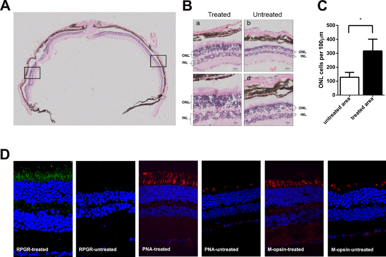
Figure 5.
Rescue of retinal structure in Rpgr−/yCas9+/WT mouse 6 months after treatment. Hematoxylin and eosin staining of retinal section of Rpgr KO mouse following vector treatment. The arrow in the left marked the injection site. (B) After treatment for 6 months, extensive and robust retinal photoreceptor preservation was observed in the treated part of the retina, with up to nine layers of rescued photoreceptors, in sharp contrast to the four layers in the untreated area in the same eye. (Upper) Magnified images of the marked areas in (A) are shown. Scale bar: 50 µm, (Lower) Magnified images of the marked areas in (A) are shown. Scale bar: 10 µm. (C) Quantitative analysis of treated and untreated part of ONL thickness from nasal-temporal retinal sections across the optic nerve head. The density of photoreceptors in the treated area was 1.5-fold more than that in the untreated part of the retina. Two-tailed paired t-test was used for statistical analysis. *P < 0.05. Error bars represent SD. INL, inner nuclear layer. (D) Restoration of Rpgr protein expression and higher numbers of PNA and M-opsin expressing cells in Rpgr−/yCas9+/WT mouse following treatment for 6 months. Rpgr are shown in green, with PNA and M-opsin staining shown in red, and nuclei are stained blue.

