Figure 7.
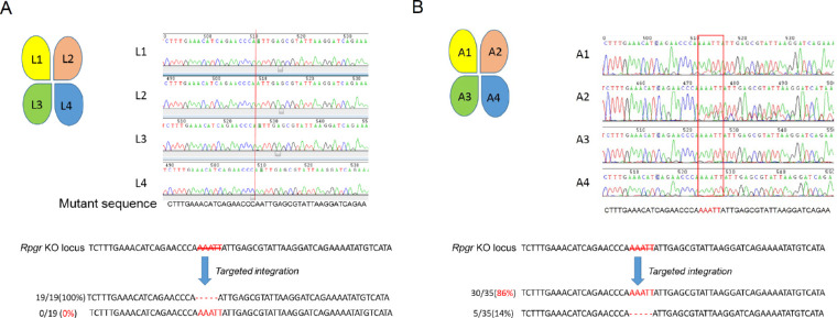
DNA sequencing analyses in Rpgr−/yCas9+/WT mouse 1 and 6 months after treatment. (A) Sequences of genomic DNA of four parts (L1, L2, L3, and L4, treated site located in L3) from treated retina 1 month after treatment. Red line shows the 5-bp deletion site. No correction was noticed. (B) Sequences of the genomic DNA of four parts (A1, A2, A3, and A4, treated site located in A3) from retina following treatment for 12 months. Red rectangle shows the nearly WT sequence in A3 and A4 parts of the retina. Sequencing analyses of clones of A3 part retina showed that 30 out of 35 clones (86%, highlighted in red) were edited correctly. Scale bar: 50 µm. INL, inner nuclear layer.

