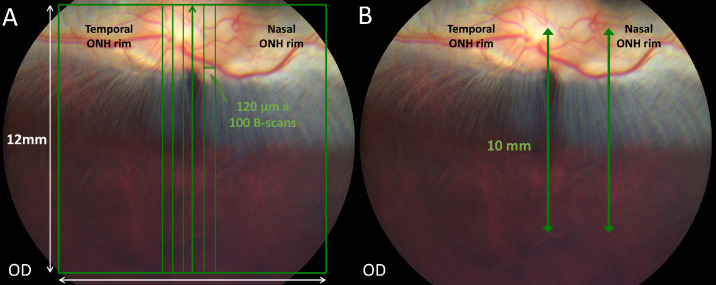
Figure 1.
SD-OCT image acquisition methodology. Color fundus pictures of a crossbred pigmented New Zealand/California rabbit. (A) A volume scan mode was used to obtain cross-sectional retinal images. The green square represents a volume scan including the visual streak ventral to the optic disc (100 vertical B-scans separated by 120 µm in a field of 12 × 12 mm). (B) A linear scan mode was used to obtain vertical line scans (10-mm scan length, 5 B-scans averaged) passing through the center of the ONH or the nasal rim of the ONH, perpendicular to the main retinal medullary rays and including the visual streak.

