Figure 4.
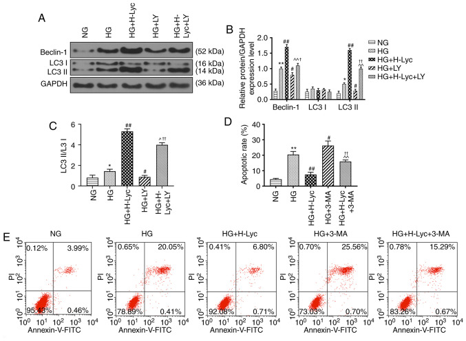
Lyc inhibits HG-induced MPC5 podocyte apoptosis by promoting autophagy via the PI3K/AKT signaling pathway. MPC5 podocytes were divided into the following groups: i) NG (5.5 mM glucose); ii) HG (25 mM glucose); iii) HG + H-Lyc (25 mM glucose + 12.5 mM Lyc); iv) HG + LY (25 mM glucose + 20 µM LY); and v) HG + H-Lyc + LY (25 mM glucose + 12.5 mM Lyc + 20 µM LY). Protein expression levels of Beclin-1, LC3I and LC3II were (A) determined by western blotting and (B) quantified. (C) The ratio of LC3II/LC3I. *P<0.05 and **P<0.01 vs. NG; #P<0.05 and ##P<0.01 vs. HG; ^P<0.05 and ^^P<0.01 vs. HG + H-Lyc; †P<0.05 and ††P<0.01 vs. HG + LY. MPC5 podocytes were divided into the following groups: i) NG (5.5 mM glucose); ii) HG (25 mM glucose); iii) HG + H-Lyc (25 mM glucose + 12.5 mM Lyc); iv) HG + 3-MA (25 mM glucose + 5 mM 3-MA); and v) HG + H-Lyc + 3-MA (25 mM glucose + 12.5 mM Lyc + 5 mM 3-MA). (D) The rate of apoptosis was determined by flow cytometry. (E) Representative scatter plots. **P<0.01 vs. NG; #P<0.05 and ##P<0.01 vs. HG; ^^P<0.01 vs. HG + H-Lyc; ††P<0.01 vs. HG + 3-MA. Lyc, lycopene; NG, normal glucose; HG, high glucose; H-Lyc, high-concentration lycopene; LY, LY294002; LC3, microtubule associated protein 1 light chain 3; 3-MA, 3-methyladenine; PI, propidium iodide.

