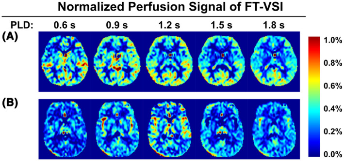
FIGURE 4.

Normalized perfusion signal images using FT‐VSI preparations acquired with PLDs from 0.6 s to 1.8 s with examples (one slice) from one female subject (31 yo) of the young‐age group (A) and one male subject (52 yo) of the middle‐age group (B)
