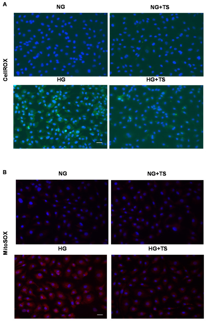
Figure 8.
Effects of Tubastatin A on cellular and mitochondrial oxidases activities in HuREC. (A) CellROX fluorescent assay showing superoxide formation (green) in HuREC exposed to HG for 48 h or to HG in the presence of 5 µM of TS (HG + TS) and compared to HuREC cultured in NG conditions in the absence (NG) or in presence of 5 µM TS (NG + TS). (B) Images of MitoSOX assay showing superoxide formation from mitochondria oxidase (red) in HuREC exposed to HG for 48 h or HG in the presence of 5 µM of TS (HG + TS), also for 48 h, and compared to HuREC cultured in NG conditions in the absence (NG) or in the presence of 5 µM TS (NG + TS). In A and B blue fluorescence show cell nuclei counterstained with DAPI. Scale bar, 50 µm.

