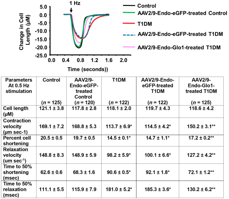
Figure 3.
Data showing evoked contractile kinetics in left ventricular myocytes isolated from control, T1DM and T1DM-treated hearts. Images above are representative evoked contraction/relation profiles of ventricular myocyte isolated from the control, AAV2/9-Endo-eGFP-treated Con, T1DM, AAV2/9-Endo-eGFP-treated T1DM, and AAV2/9-Endo-Glo1-treated DM rats field-stimulated (10 V) for 10 ms at 0.5 Hz, using a pair of platinum wires placed on opposite sides of the chamber. The extent of myocyte shortening and rates of shortening and lengthening were determined using IonWizard Version 5.0 and means ± S.E.M for n > 120 cells from n = 5 animals are shown in the table below.

