Figure 5.
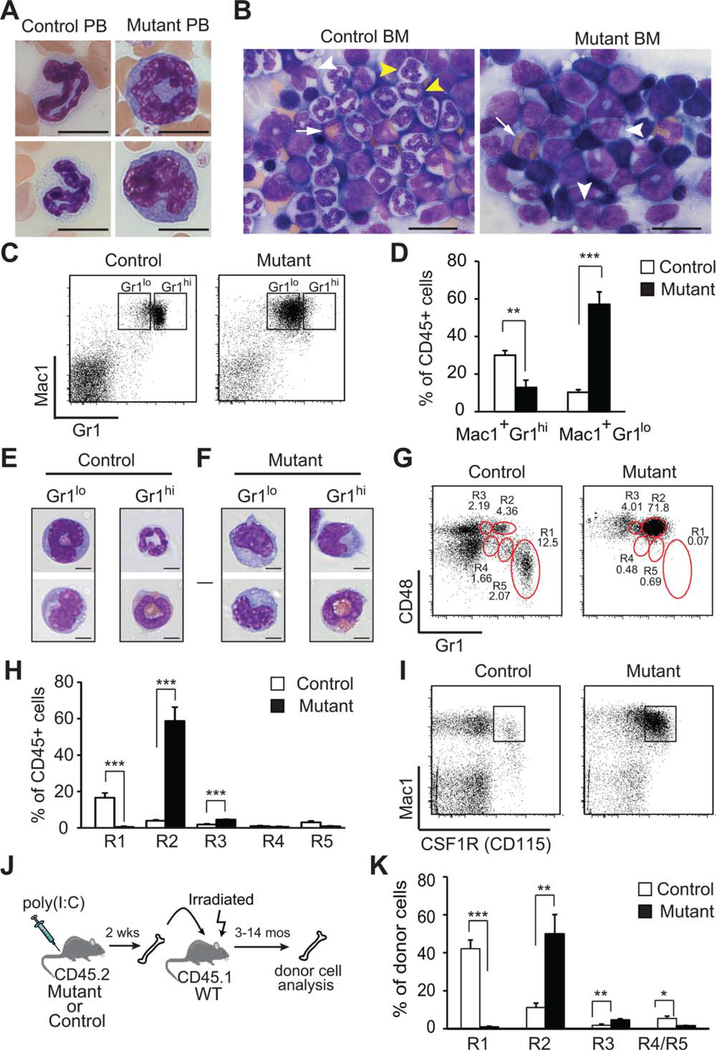
Rcor1-deficiency produces a loss of neutrophils and increased numbers of monocytes. May-Grunwald Giemsa stained (A) blood smears and (B) BM touch preparations. Maturing and mature neutrophils in PB and the BM (yellow arrow heads) were absent from Rcor1−/− mice, whereas monocytes (white arrow head) and eosinophils (white arrow) were present. (C): Mac1 and Gr1 expression on bone marrow cells. (D): A significant reduction in the proportion of Mac1+Gr1hi cells and a significant increase in the proportion of Mac1+Gr1lo cells were observed in Rcor1-deficient BM (nmutant = 9, ncontrol = 9). (E, F): Morphology of sorted myelomonocytic cells from control (E) and Rcor1-deficient (F) BM. Most Rcor1−/− cells were monocytes. (G): Phenotype of myelomonocytic cells mice based on CD48 and Gr1 expression patterns. (H): Although Rcor1-deficient mice lacked mature granulocytes (R1), they retained granulocytic precursor cells (R4, R5) and had significantly increased monocytic (R2) and bipotential (R3) cell populations relative to control mice, (nmutant = 4, ncontrol = 4). (I): Consistent with their monocytic phenotype, most Rcor1−/− Mac1+ cells coexpressed CSF1R. (J-K) Rcor1−/− donor cells maintained their abnormal monocytic phenotype after transplant into wild-type hosts (nmutant = 5, ncontrol = 5). SEM is shown; **, p < .01; ***, p < .001. Scale bars = 10 μm (A), 20 μm (B), and 5 μm (E, F). Abbreviations: BM, bone marrow; PB, peripheral blood.

