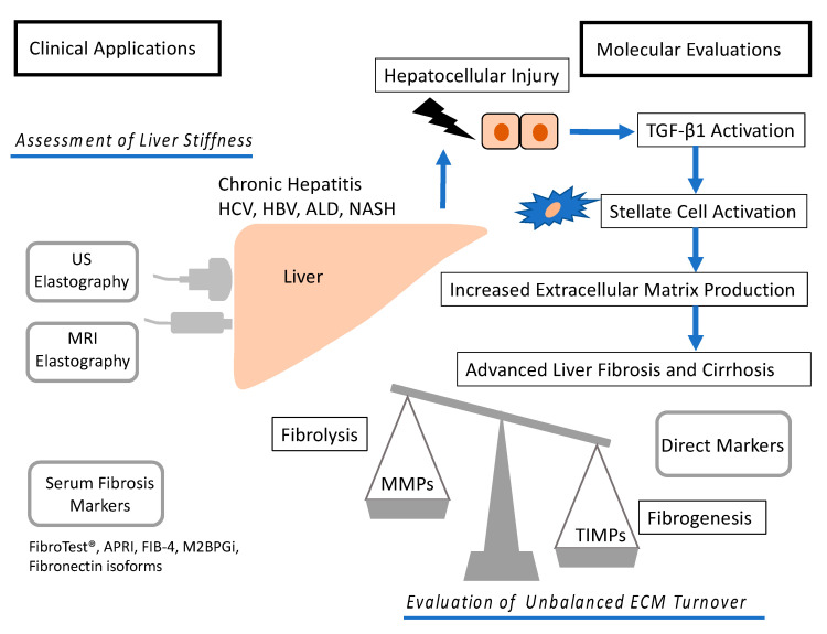
Figure 1.
Molecular mechanism and diagnosis of development of liver fibrosis. Hepatocellular injury causes TGF-β1 activation. In turn, TGF-β1 activates hepatic stellate cell and increases ECM production, causing liver fibrosis and cirrhosis. The development is depending on the balance between fibrolysis and fibrogenesis. ECM turnover is controlled by MMPs and TIMPs. Liver fibrosis can be assessed by elastography and serum markers such as aspartate aminotransferase (AST) to platelet ratio index (APRI), FIB-4, and Mac-2 binding protein glycosylation isomer (M2BPGi). US, ultrasound; MRI, magnetic resonance imaging; HCV, hepatitis C virus; HBV, hepatitis B virus; ALD, alcoholic liver disease; NASH, nonalcoholic steatohepatitis; APRI, aspartate aminotransferase to platelet ratio index; M2BPGi, Mac-2 binding protein glycosylation isomer; MMPs, matrix metalloproteinase; TIMPs, tissue inhibitors of metalloproteinases; TGF-β1, transforming growth factor β1.

