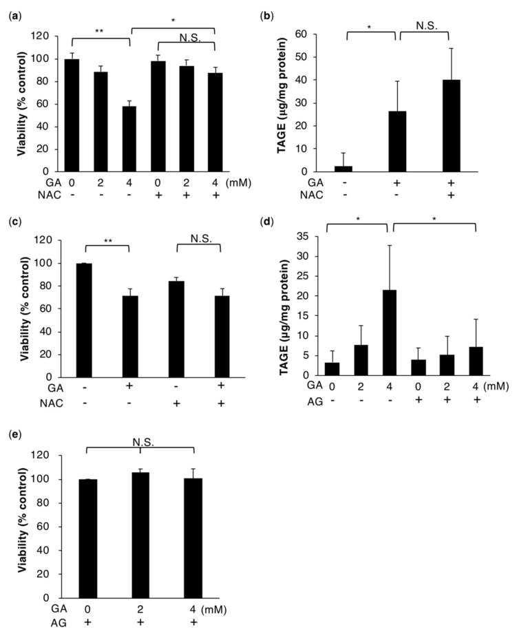
Figure 1.
TAGE accumulation-induced hepatocytes death is rescued by the antioxidant NAC. (a,c) Cell viability was measured using the CellTiter-Glo luminescent cell viability assay (n = 3). HepG2 cells (a) were treated with 0, 2, or 4 mM GA without or with 5 mM NAC for 24 h. Primary hepatocytes (c) were treated with 0 or 4 mM GA without or with 5 mM NAC for 24 h. (b,d) Slot blotting was performed to measure intracellular TAGE. (b) Cell extracts were prepared from HepG2 cells treated with 0 or 4 mM GA without or with 5 mM NAC for 24 h (n = 4). (d) Cell extracts were prepared from HepG2 cells treated with 0 or 16 mM AG for 2 h followed by 0, 2, or 4 mM GA for 24 h (n = 3). (e) HepG2 cells were treated with 16 mM AG for 2 h followed by 0, 2, or 4 mM GA for 24 h and cell viability was then measured. Results are mean ± S.D. * p < 0.05, ** p < 0.01 and N.S. (Not significant) based on a one-way ANOVA followed by Tukey’s test (a–d).

