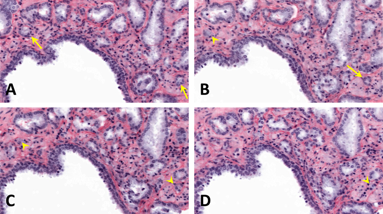
Figure 5.
Open-top light-sheet microscopy (OTLS) images of carcinoma glands that change morphology with depth. Arrows point to well-formed Gleason pattern 3 glands and arrowheads point to poorly formed Gleason pattern 4 glands. Each sequential image shows an optical section 25 microns deeper than the previous image. A, the arrows point to two well-formed glands. B, one gland is poorly formed and the other gland is well-formed. C, both glands are poorly formed at this depth. D, one gland is no longer present and the other gland is poorly formed.

