
Figure 1.
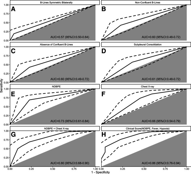
Receiver operating characteristic (ROC) curves for diagnosis of coronavirus disease 2019 pneumonitis. A–E, Lung ultrasound (LUS) patterns. F, Chest radiograph (CXR) with pneumonia and/or pulmonary edema (unilateral vs bilateral vs none). G, CXR plus LUS pattern of nondependent bilateral pulmonary edema (NDBPE; bilateral B-lines, count in superior zones ≥ inferior zones, and no pleural effusions). H, Clinical score based on room air oxygen saturation (1.5 points, ≤ 94%), temperature (1 point, ≥ 38°C), and NDBPE pattern (3 points). Gray sector indicates the ROC identity line (i.e., test no better than chance alone), and dotted line indicates the ROC 95% CI. AUC = area under the curve.