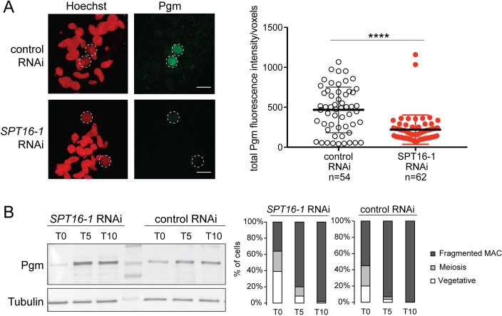
Fig 7. Spt16-1 is required for Pgm nuclear localization.
(A) Z-projections of immunolabelling with Pgm antibodies (green) and staining with Hoechst (red) in control or SPT16-1 RNAi at T5 during autogamy. Dashed white circles indicate the two developing MACs. The other Hoechst-stained nuclei are fragments from the maternal somatic MAC and the two germline MICs. Scale bar is 10 μm. Bar plots represent total Pgm fluorescence intensity in the developing new MAC divided by the number of voxels in both conditions (control or SPT16-1 RNAi). **** for p<0.0001 in a Mann-Whitney statistical test. (B) Pgm expression during development in control or SPT16-1 RNAi. Western blot of whole cell proteins extracted at T0, T5 and T10 during autogamy, when Pgm is normally detected by immunofluorescence, upon control or SPT16-1 RNAi. Pgm antibodies were used for Pgm detection and alpha tubulin antibodies for normalization. The protein ladder (PageRuler Prestained Protein Ladder, 10 to 250 kDa, ThermoFisher Scientific) is shown. Cytology of the cell population at each developmental time point is displayed in the histograms.

