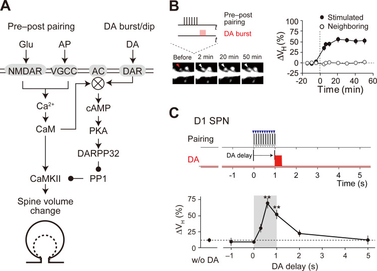
Fig 1. Reinforcement plasticity (RP) in D1 SPNs.
(A) Signaling mechanisms for RP. The indicated signals were modeled in the present study. Membreane molecules are indicated by the gray shaded areas, whereas the others are cytosolic or extracellular molecules. (B) Enlargement of stimulated spines [4]. Left: time lapse images. The target spine (red and white arrowheads) was stimulated with pre–post pairing (pairing of glutamate uncaging and action potential) and the DA burst with a 0.6-s delay (inset, also see Methods). Right: quantification of the volume change (ΔVH). From Yagishita et al., Science 26 Sep 2014:Vol. 345, Issue 6204, pp. 1616–1620 (DOI: 10.1126/science.1255514). Reprinted with permission from AAAS. (C) Critical time window for RP [4]. Top and middle: time courses of pre–post pairing (top, blue arrowheads; 10Hz, 10 times) and DA burst (middle, red lines; 30Hz, 10 times). Bottom: increase in spine volume (ΔVH) against the time delay of DA burst from the onset of pre–post pairing. From Yagishita et al., Science 26 Sep 2014:Vol. 345, Issue 6204, pp. 1616–1620 (DOI:10.1126/science.1255514). Reprinted with permission from AAAS.

